Abstract
Epidermoid cysts should be considered in the differential diagnosis for lesions causing bony erosion in the sinuses and diploic bone spaces.
Case Presentation
A 29-year old man presented with an acute onset of headache, blurred vision, and diplopia. He had no symptoms of rhinosinusitis, but a vague history of head injury in childhood. His right eye was proptotic, with markedly restricted movements superiorly and medially, but with normal vision and no chemosis. Flexible nasal endoscopy showed diffuse mucoid rhinitis. Computed tomography (CT) imaging of his sinuses was performed (Figure 1).
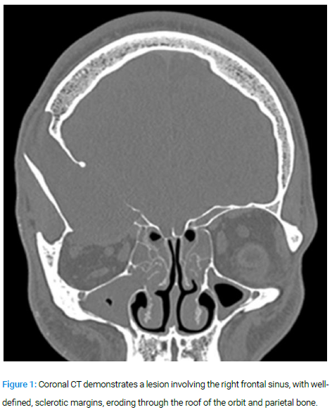
The primary differential was that of a mucocoele secondary to previous trauma to the frontal sinus. The degree of bony erosion made the exclusion of a granulomatous disease or connective tissue disorder mandatory.
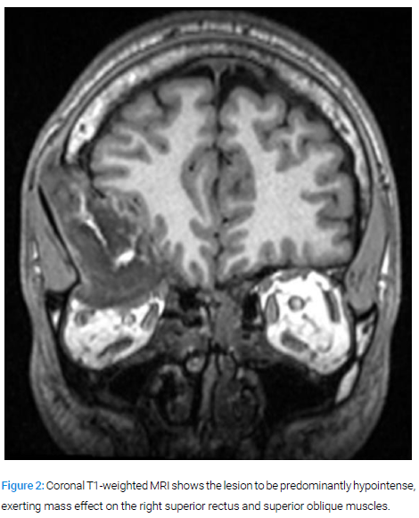
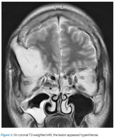
Magnetic Resonance Imaging (MRI) excluded any intradural extension (Figure 2,3).
An endoscopic transorbital superior blepharoplasty approach was used to access the lesion. Copious amounts of keratinous material, similar in appearance to that of a cholesteatoma, was found filling a bony cavity in close proximity to the right frontal sinus. The diagnosis of an epidermoid cyst was therefore made. The keratinous debris was suctioned and curetted, to remove all macroscopic disease.
Epidermoids larger than 5 cm are termed 'giant epidermoids'. Intradiploic involvement is rare but important to recognise since, as with epidermoid cysts in other sites, complete removal is essential to prevent recurrence.
Author contributions
Simon Honnet examined the patient, reviewed literature, prepared the first draft of the manuscript.
Kafui Searyoh assisted in surgery, reviewed manuscript.
Vishesh Sood interpreted CT and MRI scans, reviewed manuscript.
Darlene Lubbe performed surgery, edited final manuscript.
Keywords
Epidermoid cyst; Diplopia; Intradiploic; Bony erosion
Cite this article
Simon Honnet, Kafui Searyoh, Vishesh Sood, Darlene Lubbe. Giant intradiploic epidermoid cyst. Clin Case Rep J. 2020;1(1):1–2.
Copyright
© 2020 Simon Honnet. This is an open access article distributed under the terms of the Creative Commons Attribution 4.0 International License (CC BY-4.0).
