info@clinicalcasereportsjournal.com

Peña Ortega Maria

Department of Nephrology, San Cecilio University Hospital Spain

We cordially invite you to join our board.

Your valuable suggestions improve our journal quality. We welcome you to be part of our board members.

Fitzsimmons-Walson-Mellor Syndrome

* Peña Ortega Maria; Navas-Parejo Casado Antonio; Fernández Ortega Juan de Dios; Gómez Morales Mercedes; Caba Molina Mercedes;
  • * Peña Ortega Maria: Department of Nephrology, San Cecilio University Hospital.
  • Navas-Parejo Casado Antonio: Department of Nephrology, San Cecilio University Hospital.
  • Fernández Ortega Juan de Dios: Department of Neurology, San Cecilio University Hospital.
  • Gómez Morales Mercedes: Department of Pathology, San Cecilio University Hospital.
  • Caba Molina Mercedes: Department of Pathology, San Cecilio University Hospital.
  • Mar 06, 2020 |
  • Volume: 1 |
  • Issue: 1 |
  • Views: 2150 |
  • Downloads: 1474
  • Download PDF

Abstract

Fitzsimmons-Walson-Mellor (F-W-M) syndrome is a rare disease characterized by progressive kidney failure, sensorineural deafness, and variable spastic paraparesis.

We report the case of a 30-year-old male who had been diagnosed with non-progressive connatal encephalopathy, spastic tetraparesis of paraplegic predominance, and intellectual retardation at the age of 14 months. During adolescence, he developed proteinuria, with the subsequent detection of hypertension and elevated plasma creatinine. Analyses revealed a progressive worsening of the glomerular filtration rate, creatinine of 3.56 mg/dL, (CKD-EPI 22 mL/m), non-specific urinary sediment changes, proteinuria (1.8 g/24 h), negativity for antinuclear (ANAs) and antinuclear cytoplasmic (ANCAs) antibodies, normal C3 and C4 complements. Kidney biopsy revealed segmental and focal sclerosis lesions and severe interstitial tubular lesions.

In this complex syndrome, kidney function is affected by focal and segmental sclerotic lesions and interstitial tubule involvement. Differences in the course of kidney function among cases would reflect variations in the intensity of these lesions. Because of the very advanced nature of kidney lesions in the present patient, the biopsy findings did not offer more specific information. However, they appear consistent with the other data obtained. This condition appears to be the first case of F-W-M syndrome detected since its initial description.

Introduction

Fitzsimmons-Walson-Mellor (F-W-M) syndrome is a rare disease characterized by progressive kidney failure, sensorineural deafness, and variable spastic paraparesis. It is named after the authors who first described this syndrome in 1988 [1]. To date, only four cases have been reported of an association between F-M-W syndrome and chronic kidney disease, all in a single family with F-M-W syndrome of probable autosomal dominant heredity; its prevalence is estimated to be lower than 1/1000 000 [2].

Case Presentation

We report the case of a 30-year-old male who had been diagnosed with non-progressive connatal encephalopathy, spastic tetraparesis of paraplegic predominance, and intellectual retardation at the age of 14 months and followed up since then by our neurology department. At the age of 2 years, he had an IQ of 80 and exhibited autistic behavior, and he was diagnosed with bilateral neurosensorial hypoacusia, with hearing loss of 40% in the right ear and 50% in the left. Various fasciotomies were performed to improve his gait. At the age of 8 years, examination by pediatric nephrologists ruled out glomerular and tubular kidney disease, and electrophysiological and muscle biopsy results were normal. During adolescence, he developed proteinuria, with the subsequent detection of hypertension and elevated plasma creatinine. Analyses conducted in the adult nephrology department revealed a progressive worsening of the glomerular filtration rate, creatinine of 3.56 mg/dL, CKD-EPI of 22 mL/m, non-specific urinary sediment changes, proteinuria (1.8 g/24 h), negativity for antinuclear (ANAs)and antinuclear cytoplasmic (ANCAs) antibodies, normal C3 and C4 complements, and negative serology for HIV and hepatitis B and C. Kidney biopsy in February 2016 yielded two cylinders containing a total of 33 glomeruli with focal and segmental hyalinosis/sclerosis lesions, 26 (80%) of which were globally sclerosed. At interstitial level, 50% of tubules were atrophic, with lumina occupied by hyaline cylinders. Myointimal thickening was observed in one artery. Immunofluorescence (IF) study findings were non-specific and showed 10 glomeruli, of which 8 were sclerosed. Electron microscopy (EM) is not available at our center (Figure 1–5). Kidney function continued to progressively decline, and a hemodialysis program was initiated in May 2016.


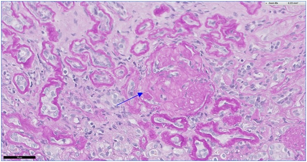
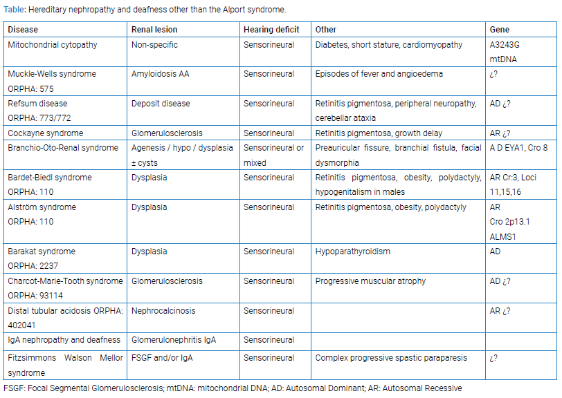
Figure 1: Global glomerulosclerosis is best appreciated in PAS stains (Arrow 40 x).


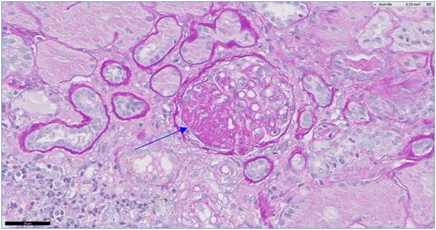
Figure 2: The affected glomerulus shows segmental sclerosing lesion (Arrow PAS 40 x).

The patient received conventional treatment with hemodialysis as well as the treatment prescribed by neurologists. He underwent cadaveric kidney transplantation in April 2017 with a good outcome.

Figure 3: Atrophic and microcystic tubules with eosinophilic casts (Arrow PAS 40 x).


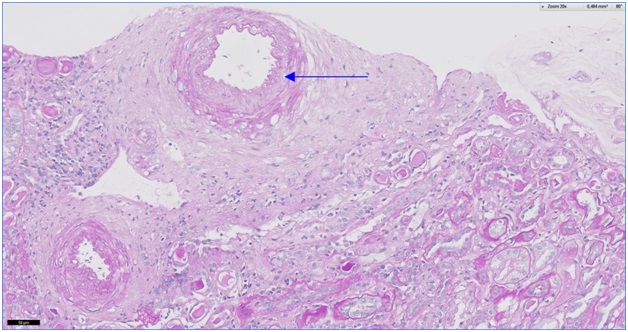
Figure 4: Intimal thickening and fibroelastosis in a small artery (PAS 20 x).


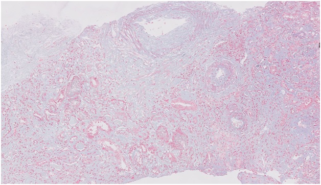
Figure 5: Trichrome stain allows more accurate assessment of interstitial fibrosis; Illustrated here is focal fibrosis with loss of tubules (Masson Trichrome 10 x).

Comments

We analyzed published data on the four generations of the family in which F-W-M syndrome was first described [1]. The index case is a male with nephropathy detected at the age of 12 years, whose kidney biopsy revealed segmental and focal glomerulosclerosis lesions and whose IF study was positive for IgA, IgG, and C3. EM showed deposits in the mesangium. His kidney function remained stable. The mother of this male developed kidney disease during her second pregnancy, with proteinuria and hypertension. A biopsy at the age of 28 years showed segmental and focal sclerotic lesions with mesangial proliferation, while IF results were positive for IgA and C3, and dense mesangial deposits were observed in the EM study. She started peritoneal dialysis at the age of 34 years. The other two cases were male and female offspring of this woman but with different fathers. They had the complete syndrome, with detection of proteinuria at the age of 8 and 6 years, respectively. No biopsies were taken. 

In the present case, kidney biopsies revealed segmental and focal sclerosis lesions and severe interstitial tubular lesions. IF results were not conclusive, probably attributable to the advanced stage of the lesions. EM study was not available.

In this complex syndrome, kidney function is affected by focal and segmental sclerotic lesions and interstitial tubule involvement, and differences in the course of kidney function among cases would reflect variations in the intensity of these lesions. Because of the very advanced nature of kidney lesions in the present patient, the biopsy findings did not offer more specific information, although they appear consistent with the other data obtained. Kidney disease and other hereditary conditions have been associated with various syndromes such as Alport syndrome, characterized by impaired renal function, hematuria, proteinuria, and ocular involvement of the lens (anterior lenticonus, cataracts) and retina [3–7]. In the attached (Table) we describe the reported hereditary nephropathy and deafness other than the alport syndrome.


Kidney disease has also been associated with deafness (e.g., IgA nephropathy [8–11], renalpolycystosis [12], membranous nephropathy [13]) and with parathyroid disorders [14,15], diabetes [16], degenerative diseases of the central nervous system [17,18] malformations [19], and hematologic disorders [20,21]. Genetic heterogeneity explains the clinical variability, given the involvement of more than 70 genes and of 14 supplementary loci where the genes have not yet been identified [22]. In the present case, metabolic diseases and tubulopathies were ruled out by pediatric nephrology test findings and myopathies by muscle biopsy results, while immunological and serological findings were normal or negative. Although little information is available on cases of F-W-M syndrome, the main pathogenic factor for kidney function prognosis appears to be the presence of segmental and focal hyalinosis lesions and severe interstitial tubule involvement. Spastic paraparesis has an incidence of 2–10/100,000 and can appear in pure or complex forms. Pure forms are characterized by a symmetric and slowly progressing spasticity of the lower extremities, while complex forms are associated with other neurological (ataxia, intellectual deficit, extra pyramidal signs, epilepsy, peripheral neuropathy) and/or extra-neurological (e.g., ichthyosis, deafness, optic atrophy, etc.) disorders. All forms of genetic transmission (autosomal dominant, recessive, “X”-linked, and mitochondrial) have been reported. SPAST (SPG4), ATL1 (SPG3), K1F5A (SPG10), and REEP1 (SPG31) genes have been implicated in 40%–50% of dominant forms and in 10%–15% of sporadic forms [22]. This variability would explain the different courses of renal function observed among members of the first reported family. F-W-M is the only syndrome associated with spastic paraparesis, nephropathy, and deafness, but it has not been related to any gene(s), possibly due to its low incidence. Our case meets the phenotypic criteria for complex progressive paraparesis, given the presence of intellectual disability, autistic traits, sensorineural deafness, and progressive nephropathy. Genetically, he can be considered to have a sporadic form of the syndrome, given the absence of a family history or consanguinity. We have been unable to trace any cases in the literature since the original report; therefore, this appears to be the first case of F-W-M syndrome detected since its initial description.

References

  1. Fitzsimmons JS, Watson AR, Mellor D, Guilbert PR. Familiar spastic paraplegia, bilateral sensorineural deafness, and intellectual retardation associated with a progressive nephropathy. J Med Genet. 1988;25(3):168–172.
  2. http://www.orpha.net/consor/cgiin/Disease_Group=2820 and Syndrome-de-Fitzsimmons-Watson-Mellor.
  3. Alport AC. Hereditary familial congenital haemorrhagic nephritis. BMJ. 1927;1:504–506.
  4. Kashtan CE. Alport syndrome. An inherited disorder of renal, ocular, and cochlear basement membranes. Medicine (Baltimore). 1999;78:338.
  5. Torra R. Síndrome de Alport y nefropatía del colágeno IV (alfa3/alfa4).  Nefrologia Suplemento Extraordinario. Pre. 2011;10904.
  6. Tryggvason K, Zhou J, Hostikka SL, Shows TB. Molecular genetics of Alport syndrome. Kidney Int. 1993;43:38.
  7. Prakash S, Chung KW, Sinha S, Barmada M, Ellis D, Ferrell RE, et al. Autosomal dominant progressive nephropathy with deafness: linkage to a new locus on chromosome 11q24.  J Am Soc Nephrol. 2003;14(7):1794–803.
  8. Ataya NL. Sensorineural deafness associated with IgA nephropathy. J Laryngol Otol. 1989;103(4):412.
  9. Bizzarri D, Duranti E, Sasdelli M, Casanova S. Familial IgA nephropathy and sensorineural deafness. Contrib Nephrol. 1990;80:13–17. 
  10. Chahin J, Ortiz A, Mendez L, Gallego E, Garcia Perez J, Garcia-Castro G, et al. Familial IgA nephropathy associated with bilateral sensorineural deafness. Am J Kidney Dis. 1992;19(6):592–596.
  11. de Serdio JL, Chahin J, Gil-Curbelo JA, Perera A, Saavedra JA. Late hereditary degenerative sensorineural hearing loss associated with IgA mesangial glomerulonephritis of probable autosomal dominant heredity. Acta Otorrinolaringol Esp. 1993;44(6):447–454.
  12. Mora C, Navarro JF, García J, Gallego E, Macía M, Méndez ML, et al. Autosomal dominant polycystic kidney disease associated with familial sensorineural deafness. Scand J Urol Nephrol. 1999;33(1):63–65.
  13. Meroni M, Volpi A, Usberti M, Battini G, Tarelli LT, Giordano F. Two brothers with idiopathic membranous nephropathy and familial sensorineural deafness. Am J Kidney Dis. 1990;15(3):269–272.
  14. Barakat AY, D’Albora JB, Martin MM, Jose PA. Familial nephrosis nerve deafness and hypoparathyroidism. J Pediatr. 1977;91(1):61–64.
  15. R.W. Bilous, G. Murty, D.B. Parkinson, R.V. Thakker, M.G. Coulthard, J. Burn. Autosomal dominant hypoparathyroidism, sensorineural deafness and renal dysplasia. New Eng J Med. 1992;327:1069–1075.
  16. Tabebi M, Mkaouar-Rebai E, Mnif M, Kallabi F, Ben Mahmoud A, Ben Saad W. A novel mutation MT-COIII m.9267G>C and MT-COI m.5913G>A mutation in mitochondrial genes in a Tunisian family with maternally inherited diabetes and deafness (MIDD) associated with severe nephropathy.  Biochem Biophys Res Commun. 2015;459(3):353–360.
  17. Feigenbaum A, Bergeron C, Richardson R, Wherrett J, Robinson B, Weksberg R. Premature atherosclerosis with photomyoclonic epilepsy, deafness, diabetes mellitus, nephropathy, and neurodegenerative disorder in two brothers: a new syndrome? Am J Med Genet. 1994;49(1):118–124.
  18. Izzi C, Maffei P, Milan G, Tardanico R, Foini P, Marshall J, et al. Familial occurrence of retinitis pigmentosa, deafness and nephropathy.  Kidney Int. 2011;79(6):691–692.
  19. Dumas R, Uziel A, Baldet P, Segond A. Glomerular lesions in the branchio-oto-renal (BOR) syndrome. Int J Pediatr Nephrol. 1982;3(2):67–70.
  20. Catarini M. Hereditary macrothrombocytopenia, deafness, nephropathy.Haematologica. 1988;73(1):43–47.
  21. Richard A, Scully C, Eveson J, Prime S. Epstein syndrome: oral lesion in a patient with nephropathy, deafness and thrombocytopenia. J Oral Pathol Med. 1991;20(10):512–513.
  22. Köhler S, Carmody L, Vasilevsky N, Jacobsen JOB, Danis D, Gourdine JP, et al. Expansion of the Human Phenotype Ontology (HPO) knowledge base and resources. Nucleic Acids Res. 2019;47(D1):D1018–D1027.

Keywords

Fitzsimmons-Walson-Mellor syndrome; Progressive kidney failure; Sensorineural deafness

Cite this article

Antonio NPC, Maria PO, Juan de Dios FO, Mercedes GM, Mercedes CM. Fitzsimmons-walson-mellor syndrome. Clin Case Rep J. 2020;1(1):1–4.

Are you interested in submitting your research paper?

Thank you for choosing us. We recommend you to go through our guidelines before submitting the paper.

Logo

Clinical Case Reports Journal is the state-of-the-art platform to showcase medical journals across scientific community and a one-stop solution for publishing the manuscripts of varied medical specialities.

Our Contacts

CLINICAL CASE REPORTS JOURNAL,
C/O Infact Publications LLC,
16192 Coastal Highway,
Lewes, Delaware 19958
USA