Clinical Image Description
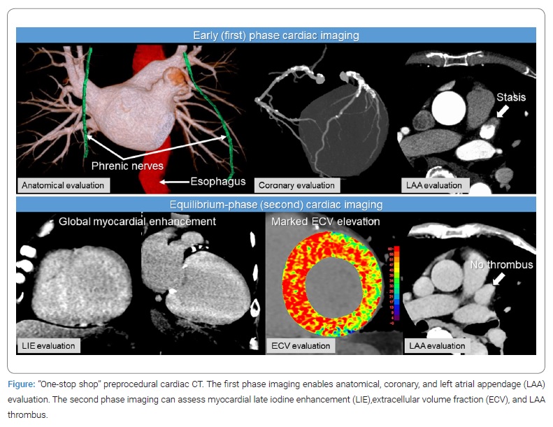
A 79-year-old man with Atrial Fibrillation (AF) underwent Computed Tomography (CT) imaging using a 320-detector row CT scanner for catheter ablation planning. A comprehensive assessment scan protocol consisting of two electrocardiography-gated cardiac acquisitions was used. The early (first) phase imaging indicated that the patient was anatomically suitable for a catheter ablation procedure and had no significant stenosis with moderate coronary artery calcification but showed a contrast-filling defect in the Left Atrial Appendage (LAA). The equilibrium (second) phase imaging showed global myocardial Late Iodine Enhancement (LIE), marked elevation in Extracellular Volume Fraction (ECV) of 72% [a normal range of 23%–28%]), and contrast fill-in in the LAA, indicating no thrombus (Figure). These findings were strongly suggestive of occult cardiac amyloidosis. The endomyocardial biopsy confirmed the presence of interstitial transthyretin amyloid deposition. Genetic testing was negative for transthyretin mutations. The patient was eventually diagnosed with AF concomitant with wild-type Transthyretin Cardiac Amyloidosis (ATTRwt-CA).

Recent studies have suggested that up to 10% to 15% of older adults with heart failure may have unrecognized ATTRwt-CA [1]. Moreover, ATTRwt-CA is often accompanied by AF. Although treatment for ATTRwt-CA was previously limited to supportive care, effective pharmacological treatments of ATTRwt-CA are now available. However, using current diagnostic strategies, it is difficult to identify the occurrence of ATTRwt-CA in AF patients before treatment.
To delineate the complex and variable anatomy of the pulmonary veins, left atrium, LAA, and surrounding structures, such as the esophagus and phrenic nerves, pre-procedure cardiac CT is widely used for ablation planning because of its accessibility, fast acquisition time, and simultaneous coronary artery evaluation. Additional equilibrium-phase cardiac imaging can allow myocardial characterization by assessing LIE and ECV [2,3]. Furthermore, it is a reasonable alternative to transesophageal echocardiography for LAA thrombus evaluation and blood stasis differentiation. This comprehensive “one-stop shop” assessment of CT imaging is believed to offer a practical and useful approach to improve the efficacy and safety of the AF catheter ablation procedure.
Conflict of Interest
The authors declare no potential conflicts of interest with respect to the research, authorship, and/or publication of this article. Informed consent was obtained for this publication.
Keywords
Cardiac amyloidosis; Computed tomography; Catheter ablation; Atrial fibrillation
Cite this article
Emoto T; Oda S; Kidoh M; Hayashi H; Sasaki G; Hokamura M, et al. Occult cardiac amyloidosis:“One-stop shop” preprocedural cardiac computed tomography on catheter ablation in atrial fibrillation. Clin Case Rep J. 2023;4(1):1–2.
Copyright
© 2023 Seitaro Oda. This is an open access article distributed under the terms of the Creative Commons Attribution 4.0 International License (CC BY-4.0).

