A Case Report of Retained Sea Urchin Spines Requiring Surgical Removal
* Dora LE;
-
* Dora LE: Department of Internal Medicine, Medvadis AG–Limmattal, Urdorf, Switzerland
Abstract
Background: Sea urchin injuries commonly occur in marine environments and often resolve without intervention. Retained spines can cause persistent pain, localized inflammation, and soft tissue infection. We present a case requiring surgical management.
Case Presentation: A 30-year-old healthy man presented with persistent pain in his right hand and both feet one week after stepping on and contacting a sea urchin. The patient attempted self-removal of the spines; however, several fragments remained embedded. Clinical examination revealed puncture wounds with mild erythema and tenderness, and no systemic signs of infection were observed. Surgical exploration under general anesthesia allowed the successful removal of the spines and thorough debridement.
Methods: Surgical exploration of the right hand and both feet revealed multiple retained sea urchin spine fragments, which were excised. The wounds were then irrigated and left partially open. Subsequently, the patient received a 5-day course of amoxicillin–clavulanate and postoperative local wound care.
Results: Healing was uneventful. At follow-up, the patient had no residual pain or infection, and full function of the hand and feet was observed.
Conclusion: This case underscores the importance of clinical suspicion for retained spines and the role of surgery when conservative management fails. Early surgical intervention may prevent complications and achieve excellent outcomes.
Introduction
Sea urchin injuries commonly affect swimmers and divers in warm coastal regions. The spines are sharp, brittle, and mainly composed of calcium carbonate. Most injuries resolve on their own, but retained fragments can cause soft-tissue inflammation, infection, or tenosynovitis [1].
Diagnosis frequently relies on clinical suspicion, as the spines are radiolucent and often not visible on standard X-rays. Consequently, in symptomatic cases, especially when localized infection or functional impairment is evident, surgical removal becomes necessary.
Building on these considerations, we present a case of multiple retained sea urchin spines involving the hand and feet of a healthy young adult that required operative management.
Case Presentation
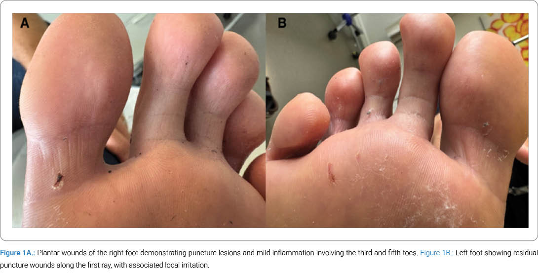
A 30-year-old male patient presented with pain and swelling involving the right palm, fingers, and plantar surfaces of both feet one week after stepping on a sea urchin while swimming barefoot (Figure 1A, Figure 1B). The patient also reported reflexively grasping the sea urchin, resulting in additional hand injuries.
Following his initial injury, the patient attempted to remove the spines using tweezers and home remedies. Despite partial improvement, he continued to experience persistent localized pain and progressively increasing tenderness at multiple puncture sites.
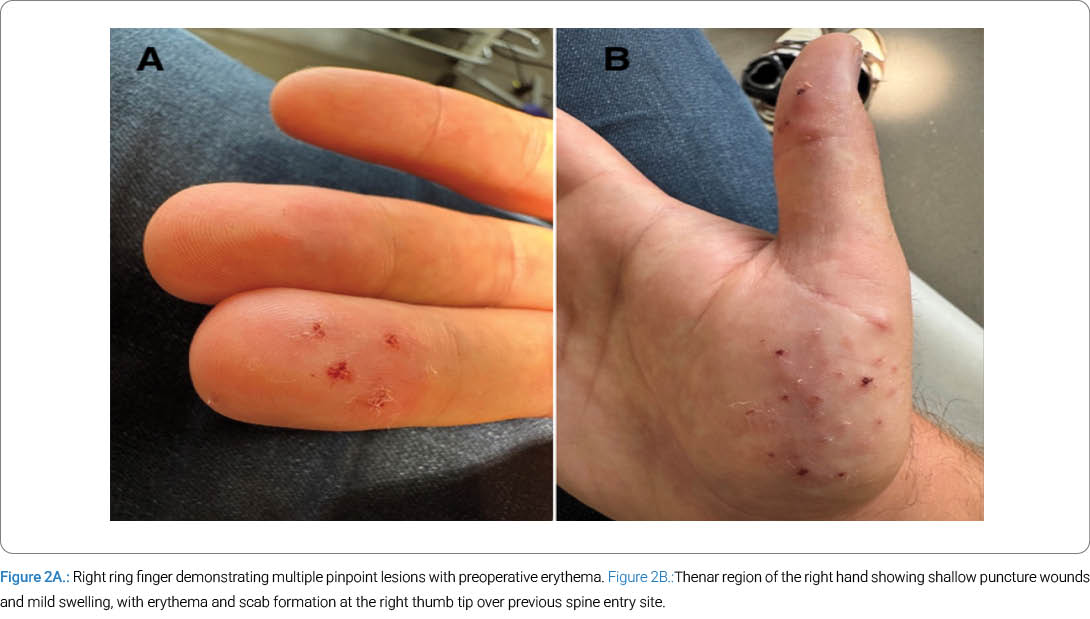
Upon further assessment, examination revealed puncture wounds in the thenar region (Figure 2B), the distal phalanges of the thumb and ring finger (Figure 2A), the right heel, and the forefoot (first, third, and fifth rays). Localized erythema and mild swelling were present; however, no systemic signs of infection were evident.
Given the patient’s ongoing symptoms and the suspicion of retained spines, surgical exploration was indicated.
Surgical Procedure
Under general anesthesia, surgical exploration was performed on the right hand and both feet. A pneumatic tourniquet set at 250 mmHg was used on the right arm. All discernible sea urchin spine fragments were carefully removed, after which the affected areas were debrided and thoroughly irrigated with sterile saline [2].
Larger wounds were left partially open to promote drainage, and sterile dressings were applied.
Postoperatively, the patient received:
- Amoxicillin–clavulanate 625 mg three times daily for 5 days.
- Weight-adjusted enoxaparin prophylaxis.
- Instructions to keep the wounds clean and dry.
Outcome
The patient was reviewed on postoperative days 3 and 14. At each visit, all wounds demonstrated satisfactory healing, with no evidence of infection, discharge, or retained fragments. Furthermore, the patient exhibited a full range of motion of the hand with no sensory deficits [3]. No scar hypertrophy or functional impairment was observed during these assessments.
Discussion
Although most sea urchin injuries heal on their own, retained spines can cause persistent symptoms or complications [4].
Common complications include:
- Local soft tissue inflammation.
- Secondary bacterial infection.
- Foreign body granulomas.
- Chronic pain or tenosynovitis.
Identifying retained sea urchin spines can be challenging [5]. Due to their radiolucent nature, they are often difficult to detect on standard X-rays. As a result, clinical suspicion is essential, particularly in patients presenting with persistent pain or localized swelling following exposure [6]. Surgical exploration should be considered under the following conditions [7]:
- Failure of conservative management.
- Persistent pain or functional impairment.
- Development of infection.
Conclusion
This case shows that minor sea urchin injuries can progress to persistent foreign body reactions and localized inflammation. When clinical suspicion is high and conservative management fails, surgical removal is a safe, effective intervention. Early management helps prevent chronic pain and functional impairment.
Acknowledgments
We thank everyone who supported us during the preparation of this case report.
Patient Perspective: “I didn’t expect something so small to cause such a big problem. Trying to remove the spines myself only made the pain worse. Surgery helped a lot, and now everything is back to normal.”
Consent: Written informed consent was obtained from the patient for publication of this case report and the use of clinical images.
Funding: No external funding was received for this case report.
Conflicts of Interest
The author declares no potential conflicts of interest with respect to the research, authorship, and/or publication of this article. Informed consent was obtained for this publication.
References
- Gulati A, Maheshwari R, Kaushal R. Sea urchin injuries: A case report and review of management. J Orthop Case Rep. 2010;1(2):27–29.
- Heggie TW, Press DJ. Sea urchin injuries. Wilderness Environ Med. 2010;21(3):261–267.
- Auerbach PS. Marine envenomations. N Engl J Med. 1991;325(7):486–493.
- Thomas I, Lahlou Z, Brutus JP. Sea urchin spine injuries of the hand: A report of two cases and literature review. Hand Surg Rehabil. 2016;35(4):307–309.
- Yoshino K, Teranishi S, Fujita Y. Surgical treatment for chronic tenosynovitis due to sea urchin spine: A case report. J Hand Surg Eur Vol. 2013;38(7):774–775.
- Zafren K, Thurman R, Jones ID. Sea Urchin Envenomation. In: Knoop KJ, Stack LB, Storrow AB, Thurman R. editors. The Atlas of Emergency Medicine: McGraw-Hill; 2021.
- Dahl WJ, Jebson P, Louis DS. Sea urchin injuries to the hand: a case report and review of the literature. Iowa Orthop J. 2010;30:153–156.
Cite this article:
Dora LE. A case report of retained sea urchin spines requiring surgical removal. Clin Case Rep J. 2026;7(3):1–3.
Keywords
Sea urchin; Foreign body; Foot injury; Hand trauma; Surgical debridement
Copyright
© 2026 Lilla Edit Dora. This is an open access article distributed under the terms of the Creative Commons Attribution 4.0 International License (CC BY-4.0).
