Acute Coronary Syndrome Secondary to Type I Kounis Syndrome Following Suspected Wheat Hypersensitivity
* Prashant J;
Sambaji AS;
-
* Prashant J: Department of Critical Care Medicine, Manipal Hospitals, India
-
Sambaji AS: Department of Cardiology, Manipal Hospitals, India
Abstract
Kounis syndrome (KS) is an acute coronary syndrome occurring in association with a hypersensitivity reaction. Type I variant is characterized by allergy-mediated coronary vasospasm in patients without pre-existing obstructive coronary artery disease. We describe a 31-year-old man who presented with diffuse pruritic urticaria after ingesting bread prepared from refined wheat flour. He had no conventional cardiovascular risk factors and reported previous cutaneous reactions to wheat consumption. During treatment of the allergic episode, he developed abrupt palpitations, diaphoresis, severe chest pain, tachycardia, and hypotension. Electrocardiography demonstrated ST-segment depression across leads V1-V6, and serum troponin I was elevated to 1.277 ng/mL. The ischemic electrocardiographic abnormalities resolved within one hour as his hemodynamic status improved. Echocardiography showed anterior wall hypokinesia with mild left ventricular systolic dysfunction and an ejection fraction of 50%. Coronary angiography performed 24 hours after symptom onset demonstrated normal epicardial coronary arteries without significant stenosis. The concurrence of a systemic allergic presentation, objective evidence of transient myocardial ischemia, and unobstructed coronary arteries supported the diagnosis of Type I Kounis syndrome. This case emphasizes the need to consider an allergy-mediated coronary event when acute myocardial ischemia occurs in a young patient with concomitant hypersensitivity manifestations and no established Coronary Artery Disease (CAD).
Abbreviations
ACS: Acute Coronary Syndrome; CAD: Coronary Artery Disease; ECG: Electrocardiogram; EF: Ejection Fraction; KS: Kounis Syndrome; LV: Left Ventricular
Introduction
Kounis Syndrome (KS) describes acute myocardial ischemia that occurs in the setting of an allergic or hypersensitivity reaction. Coronary manifestations are attributed to the cardiovascular effects of inflammatory mediators released during mast cell activation and platelet stimulation, including histamine, leukotrienes, tryptase, cytokines, and platelet-activating factor. These mediators can alter the coronary vascular tone and, in susceptible patients, may also contribute to plaque destabilization or thrombosis [1,2].
Three clinical phenotypes have been identified. Type I KS occurs in patients without established obstructive coronary artery disease and is predominantly mediated by coronary vasospasms. Type II occurs in the presence of underlying atherosclerotic disease, where the allergic response may precipitate vasospasm and/or acute plaque-related events. Type III refers to hypersensitivity-associated stent thrombosis [1,3]. The syndrome has been described after exposure to medications, foods, insect stings, vaccines, and other allergens; however, its true frequency remains uncertain because recognition depends heavily on the clinical context [3,4].
The diagnostic challenge is greater when transient electrocardiographic abnormalities and biomarker elevations resemble those of conventional Acute Coronary Syndrome (ACS). In such circumstances, the temporal relationship between hypersensitivity manifestations and myocardial ischemia, together with subsequent assessment of coronary anatomy, becomes central to diagnosis. We report a case of a young man with a wheat-associated allergic episode followed by hemodynamic instability, reversible ischemic electrocardiographic changes, troponin elevation, and angiographically normal coronary arteries.
Case Presentation
A 31-year-old man presented to the Emergency Department of Manipal Hospital, Whitefield, with a 12-hour history of intensely pruritic cutaneous eruptions involving both upper limbs. The symptoms began after he consumed bread prepared from refined wheat flour the night before. He was a non-smoker with no recognized coronary risk factors and reported no family history of coronary artery disease. A food allergy panel performed on May 12, 2022, was previously reported as positive for chicken and crab. In addition, he described intermittent episodes of arm rash after consuming wheat-containing foods.
At presentation, he was fully alert, with a Glasgow Coma Scale score of 15/15. His temperature was 98 °F, heart rate was 80 beats/ min, blood pressure was 130/80 mmHg, and respiratory rate was 20 breaths/min. Jugular venous pressure was not elevated, and peripheral edema was absent. Examination demonstrated diffuse urticarial lesions over the upper and lower extremities. Cardiac auscultation revealed normal first and second heart sounds, with no audible third or fourth heart sounds. Air entry was equal bilaterally.
The treating team considered an acute hypersensitivity reaction and administered intravenous pheniramine maleate 1 mL slowly together with hydrocortisone 100 mg. Shortly thereafter, the patient developed sudden palpitations, dizziness, a sensation of head heaviness, severe chest pain, and sweating. At that time, his heart rate had increased to 170 beats/min, blood pressure had fallen to 85/50 mmHg, and respiratory rate was 27 breaths/min. Intravenous crystalloid resuscitation was initiated with a 1,500 mL normal saline bolus, followed by an infusion at 100 mL/h. Intravenous ranitidine was also administered. A 12-lead electrocardiogram obtained during the episode showed ST-segment depression in the anterior precordial leads V1-V6 (Figure 1).
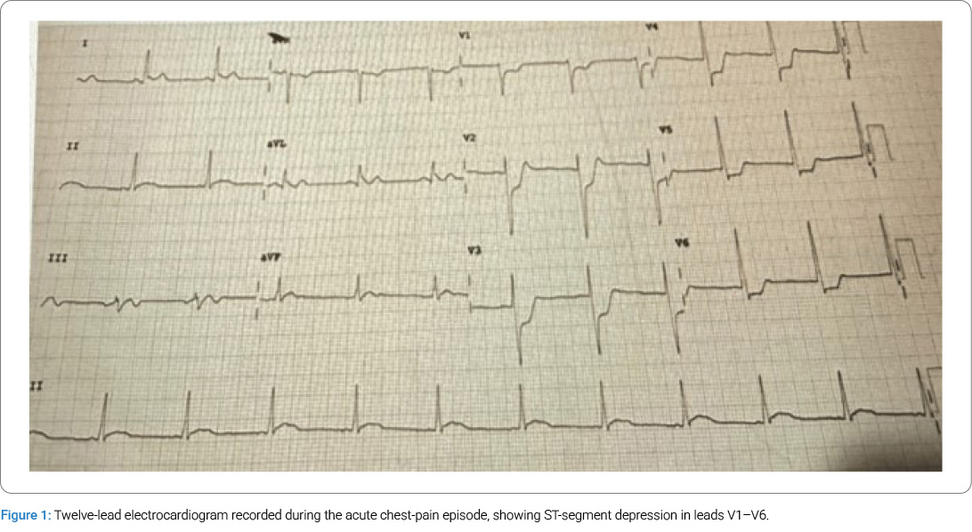
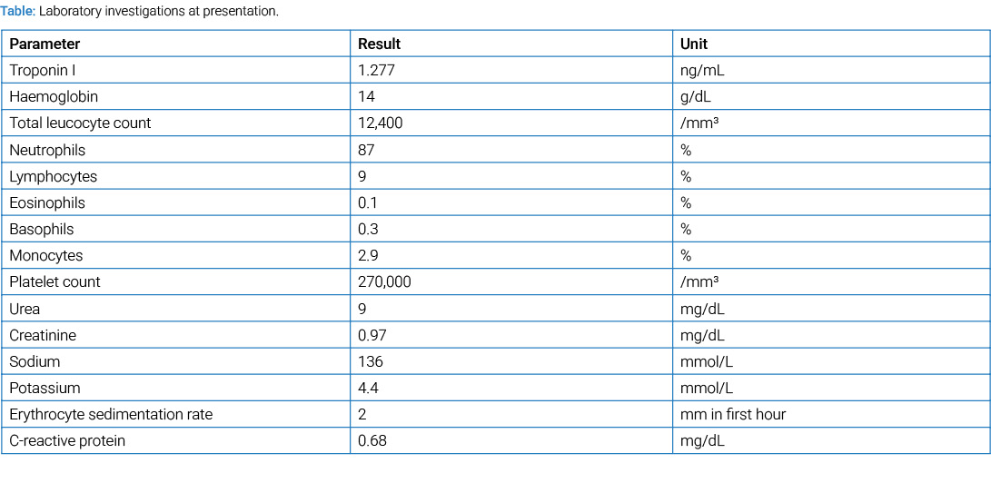
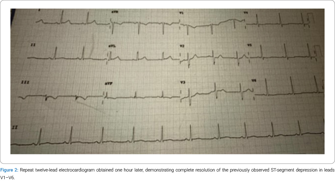
Serum troponin I levels were elevated to 1.277 ng/mL. The total leukocyte count was 12,400/mm3 with neutrophil predominance (87%). The remaining laboratory findings are presented in Table. Aspirin 325 mg was administered orally as antiplatelet therapy. Clinical improvement was accompanied by normalization of the heart rate to 75 beats/min and blood pressure of 100/60 mmHg. Repeat electrocardiogram one hour after the initial tracing demonstrated complete resolution of the previously observed ST-segment depression (Figure 2).
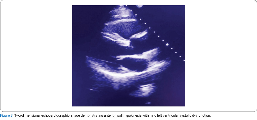
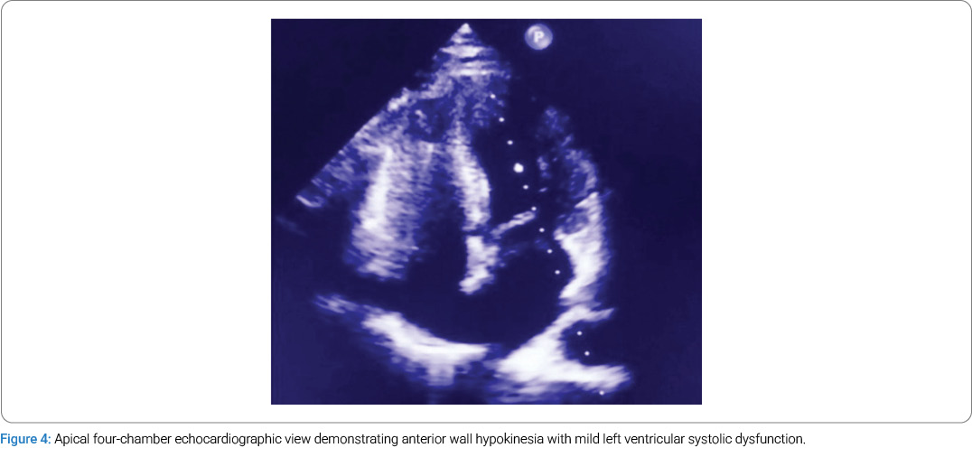
Two-dimensional echocardiography demonstrated anterior wall hypokinesia with mild left ventricular systolic dysfunction; the estimated left ventricular ejection fraction was 50% (Figure 3, Figure 4). In view of the concurrent allergic manifestations, dynamic ischemic electrocardiographic abnormalities, elevated troponin, and regional wall motion abnormality, an allergy-associated ACS due to coronary vasospasm was considered. The patient was transferred to a high-dependency unit for continued observation and treatment.
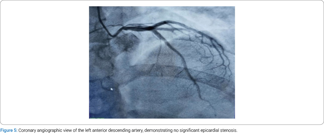
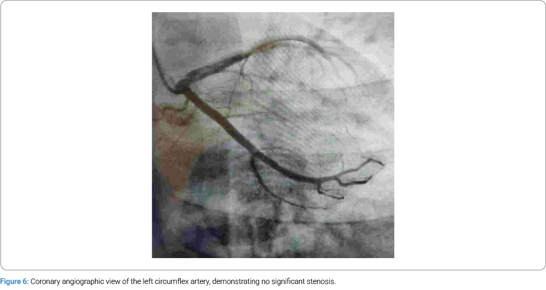
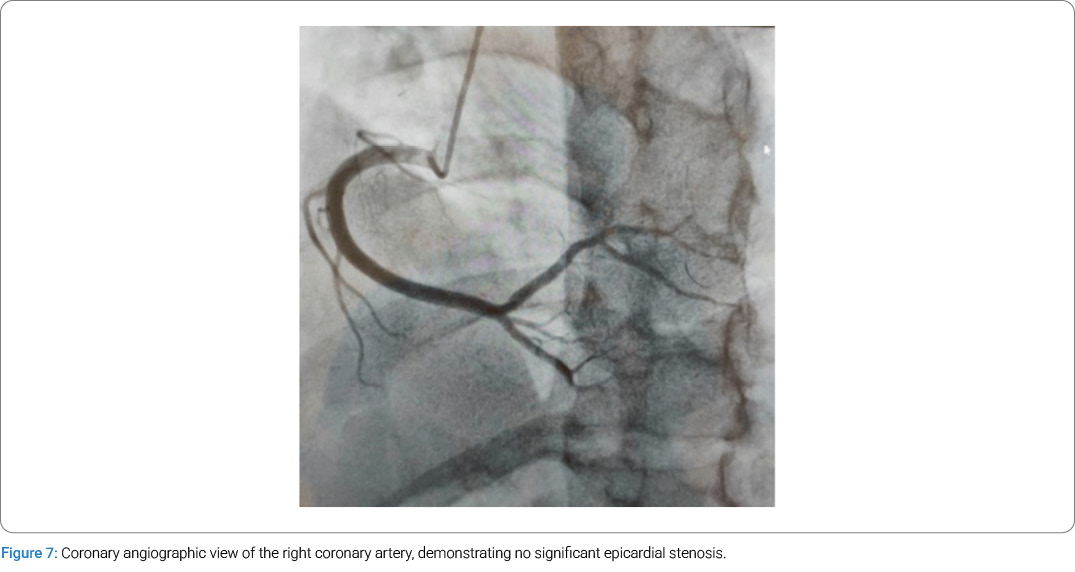
Dual antiplatelet therapy, statin therapy, and low-molecular-weight heparin were continued during the inpatient monitoring period. Coronary angiography performed approximately 24 hours after onset of the cardiac symptoms demonstrated normal epicardial coronary arteries, with no significant stenosis in the left anterior descending, left circumflex, or right coronary arteries (Figure 5–Figure 7). In the absence of obstructive coronary disease, the overall clinical pattern was considered to be most consistent with Type I Kounis syndrome.
No further episodes of chest pain, hemodynamic instability, or allergic symptoms occurred during hospitalization. The patient was subsequently transferred to the general ward and discharged the following day on antiplatelet, statin, and antianginal therapy with advice for close outpatient review. At the one-week follow-up, he remained clinically well, without recurrent chest pain or allergic manifestations.
Discussion
The principal diagnostic feature of this case was the close clinical association between an acute hypersensitivity presentation and objectively documented myocardial ischemia in a patient without obstructive coronary disease. Although urticaria preceded the cardiac symptoms, the subsequent episode was accompanied by marked tachycardia and hypotension, transient ST-segment depression, a positive troponin I result, and anterior regional wall motion abnormality. These findings suggest myocardial involvement rather than an isolated allergic reaction. The later demonstration of normal epicardial coronary arteries makes fixed atherosclerotic obstruction an unlikely explanation for the acute ischemic episode.
This constellation is compatible with Type I KS. In this phenotype, vasoactive mediators released during the allergic response can produce transient coronary constriction in angiographically normal vessels [1,2]. A normal angiogram obtained after symptom resolution does not exclude an earlier vasospastic event, as the defining abnormality may be dynamic rather than structural. Similar reports have described reversible electrocardiographic changes and normal or non-obstructed coronary arteries after allergy-associated myocardial ischemia [2,5]. In the present patient, rapid normalization of the electrocardiogram within one hour further supports the transient coronary vasomotor mechanism.
Alternative explanations were also considered in this study. Conventional plaque-rupture acute coronary syndrome was less likely because the patient was young, lacked recognized coronary risk factors, and had angiographically normal epicardial arteries. Myocarditis and stress cardiomyopathy can present with chest pain, troponin elevation, and transient ventricular dysfunction, but neither readily explains the close temporal association with urticaria, abrupt hemodynamic compromise, and rapidly reversible ischemic ST-segment changes. Coronary spasm provocation and cardiac magnetic resonance imaging were not performed; therefore, these competing diagnoses cannot be completely excluded. Nevertheless, the sequence of hypersensitivity symptoms followed by transient myocardial ischemia, together with the recovery of the electrocardiogram and the absence of obstructive coronary disease, strongly favors an allergy-mediated vasomotor event. This diagnostic reasoning is clinically important because Kounis syndrome may be overlooked when cardiac manifestations dominate the presentation. Conversely, attributing myocardial injury to allergy alone without objective cardiac assessment can delay the recognition of true acute coronary syndrome. Therefore, the parallel evaluation of the allergic trigger, serial electrocardiograms, biomarkers, ventricular function, and coronary anatomy is central to appropriate classification [1–4].
Wheat was considered the probable precipitating exposure because the cutaneous eruption followed ingestion of refined wheat-flour bread, and the patient recalled previous episodes of rash after consuming wheat. Food allergens are recognized triggers of KS, and food-associated cases have been reported in patients without a history of coronary artery disease [1,5]. Nevertheless, the strength of causal attribution should be interpreted with caution. The available records do not document wheat-specific immunoglobulin E, skin prick testing, serum tryptase levels during the acute event, or a supervised food challenge. Furthermore, cardiovascular deterioration occurred after the administration of pheniramine and hydrocortisone, creating temporal complexity in assigning a single trigger. Therefore, this case supports the hypothesis that a clinically suspected wheat-associated hypersensitivity reaction, rather than a laboratory-confirmed wheat allergy, is the proximate cause.
The management of KS requires simultaneous consideration of the allergic process and coronary syndrome. This can be difficult because the relative contributions of vasospasm, thrombosis, and hemodynamic compromise vary among patients, and therapeutic priorities may differ across KS phenotypes [1,3,4]. In Type I disease, resolution of the hypersensitivity response and coronary spasm may be followed by rapid normalization of ischemic changes. In contrast, patients with Type II or Type III disease may require treatment directed at plaque-mediated ACS or stent thrombosis. Published case series and reviews emphasize individualized management based on hemodynamic status, electrocardiographic evolution, biomarker findings, and coronary anatomy, rather than a single, uniform protocol [2–4].
This study has several limitations. Serum tryptase and other markers of mast cell activation were not reported, the suspected wheat trigger was not confirmed by targeted allergy testing, and no provocative coronary spasm testing was performed. Only short-term follow-up at one week was available. These limitations do not negate the observed concurrence of hypersensitivity and myocardial ischaemia, but they restrict certainty regarding the precise allergen and the mechanistic confirmation of vasospasm. The diagnostic value of this case lies in the integrated pattern of allergic manifestations, transient ischemic electrocardiographic changes, myocardial injury, regional ventricular dysfunction, and unobstructed epicardial coronary arteries.
Conclusions
Type I Kounis syndrome should be included in the differential diagnosis when objective evidence of acute myocardial ischemia develops during or shortly after a hypersensitivity reaction, particularly in younger patients without conventional coronary risk factors. In this patient, reversible ST-segment depression, troponin elevation, transient left ventricular dysfunction, and normal coronary angiography findings supported an allergy-mediated vasospastic mechanism. Recognition of this clinical association can facilitate the prompt evaluation of both the allergic and cardiac components of the presentation. Where the inciting allergen has not been confirmed by targeted testing, causal attribution should remain appropriately cautious and conservative.
Conflict of Interest
The authors declare no potential conflicts of interest with respect to the research, authorship, and/or publication of this article. Informed consent was obtained for this publication.
References
- Kounis NG. Kounis syndrome: an update on epidemiology, pathogenesis, diagnosis and therapeutic management. Clin Chem Lab Med. 2016;54(10):1545–1559.
- Li J, Zheng J, Zhou Y, Liu X, Peng W. Acute coronary syndrome secondary to allergic coronary vasospasm (Kounis Syndrome): a case series, follow-up and literature review. BMC Cardiovasc Disord. 2018;18(1):42.
- Benchea LC, Anghel L, Scripcariu DV, Diaconu A, Zanfirescu RL, Lucaci LV, et al. Kounis syndrome in clinical practice: insights from clinical case series and mechanistic pathways. J Clin Med. 2025;14(3):768.
- Rochel-Perez E, Santaularia-Tomas M, Martin-Dorantes M, Villareal-Jimenez E, Olivera-Mar A, Sanchez-Felix E, et al. Triggers, types, and treatments for Kounis syndrome: a systematic review. Clin Pract. 2025;15(3):59.
- Rodríguez-Ruiz C, Puig-Carrion GD, Delgado-Nieves A, López-Candales A. Kounis syndrome: a more commonly encountered cause of acute coronary syndrome. Heart Views. 2019;20(3):122–125.
Cite this article
Prashant J, Sambaji AS. Acute Coronary Syndrome Secondary to Type I Kounis Syndrome Following Suspected Wheat Hypersensitivity. Clin Case Rep J. 2026;7(4):1–6.
Keywords
Kounis syndrome; Acute coronary syndrome; Coronary vasospasm; Hypersensitivity; Wheat hypersensitivity
Copyright
© 2026 Prashant J. This is an open access article distributed under the terms of the Creative Commons Attribution 4.0 International License (CC BY-4.0).