Abstract
Coccidioidomycosis, colloquially known as “Valley fever,” is a fungal infection caused by Coccidioides immitis or Coccidioides posadasii. Exposure typically occurs through inhalation of airborne spores from disturbed soil or dust in endemic regions of the southwestern United States. The disease most commonly presents as a self-limited flu-like illness or community-acquired pneumonia. However, it may also manifest with extrapulmonary involvement, including meningitis, erythema nodosum, arthralgias, and osteolytic bone lesions. We report a case of disseminated coccidioidomycosis in a young, previously healthy patient, with osteolysis in the vertebral column with spread to the Central Nervous System (CNS) causing meningitis, further complicated by treatment-resistant hydrocephalus.
Introduction
Coccidioidomycosis is a dimorphic fungal infection, endemic to the Southwestern United States, that is typically caused by inhalation of spores in the soil. In endemic regions, coccidioidomycosis accounts for 25% of community-acquired pneumonia cases [1]. Coccidioidomycosis presents with a wide spectrum of manifestations, ranging from asymptomatic infection to respiratory illness and extrapulmonary disease. Disseminated disease occurs in approximately 0.5%–2% of cases and may involve the CNS, bones, joints, lymph nodes, and skin [2]. Primary pulmonary coccidioidomycosis commonly presents with symptoms including cough, fever, pleuritic chest pain, fatigue, and night sweats. Extrapulmonary manifestations of coccidioidomycosis most commonly include osteoarticular involvement or vertebral infection. Coccidioidal meningitis commonly presents with symptoms including headache, photophobia, nausea, vomiting, and altered mental status. Complications of coccidioidal meningitis include hydrocephalus, vasculitis, cerebral ischemia and infarction, vasospasm, and hemorrhage. Hydrocephalus frequently necessitates ventriculoperitoneal shunt placement [3]. Immunocompromised individuals are at increased risk for infection or severe disseminated disease.
Additionally, certain ancestral backgrounds, including African and Filipino ancestry, are associated with a higher risk of severe disease [3]. Diagnosis typically involves chest X-ray along with Computed Tomography (CT) or Magnetic Resonance Imaging (MRI), depending on symptoms, and lumbar puncture among those experiencing symptoms suspicious for coccidial meningitis. Lumbar puncture in the setting of coccidioidal meningitis may show low-grade lymphocytic pleocytosis, elevated protein, and low-normal glucose [2]. Coccidioidomycosis is most commonly diagnosed through serologic testing, with Enzyme Immunoassay (EIA) of IgM or IgG antibodies used for initial screening and confirmatory testing with immunodiffusion complement fixation required due to limited specificity of EIA. Definitive diagnosis relies on histopathology of samples obtained from involved sites, whereas other modalities, such as antigen detection and PCR, may facilitate earlier diagnosis [2]. Treatment typically consists of azoles and amphotericin in severe or disseminated cases.
Case Presentation
A previously healthy 22-year-old Hmong male residing in the southwestern United States presented to the Emergency Department (ED) with headache, nausea, and vomiting. He denied any history of immunosuppressive therapy or recreational drug use. On presentation, he was afebrile and hemodynamically stable. Initial evaluation, including Chest Radiography (CXR), influenza testing, and SARS-CoV-2 testing, was negative, and he was diagnosed with a viral upper respiratory infection.
The following day, he returned to the ED with a worsening headache and recurrent vomiting. Vital signs remained stable and neurologic examination revealed no focal deficits. Noncontrast CT (NC-CT), contrast-enhanced CT, and CT angiography of the head were unremarkable. Epstein–Barr Virus (EBV) serology was positive for IgG and repeat respiratory viral testing was negative. Symptoms were attributed to a viral illness with superimposed migraine.
Over the following week, the patient participated in two telephone encounters. During the initial telephone encounter, he described his headache as sinus pressure exacerbated by leaning forward. He also complained of multiple episodes of vomiting, nausea, diarrhea, subjective fever and chills, upper abdominal pain, and decreased urine output. The patient was prescribed ondansetron and amoxicillin-clavulanate for presumed bacterial sinusitis. During the second telephone encounter, the patient noted progressive headache, new-onset photophobia, lightheadedness, intermittent subjective fever and chills, decreased urine output, persistent vomiting, and inability to tolerate antibiotics. These symptoms were accompanied by an approximately 6.8 kg weight loss since symptom onset.
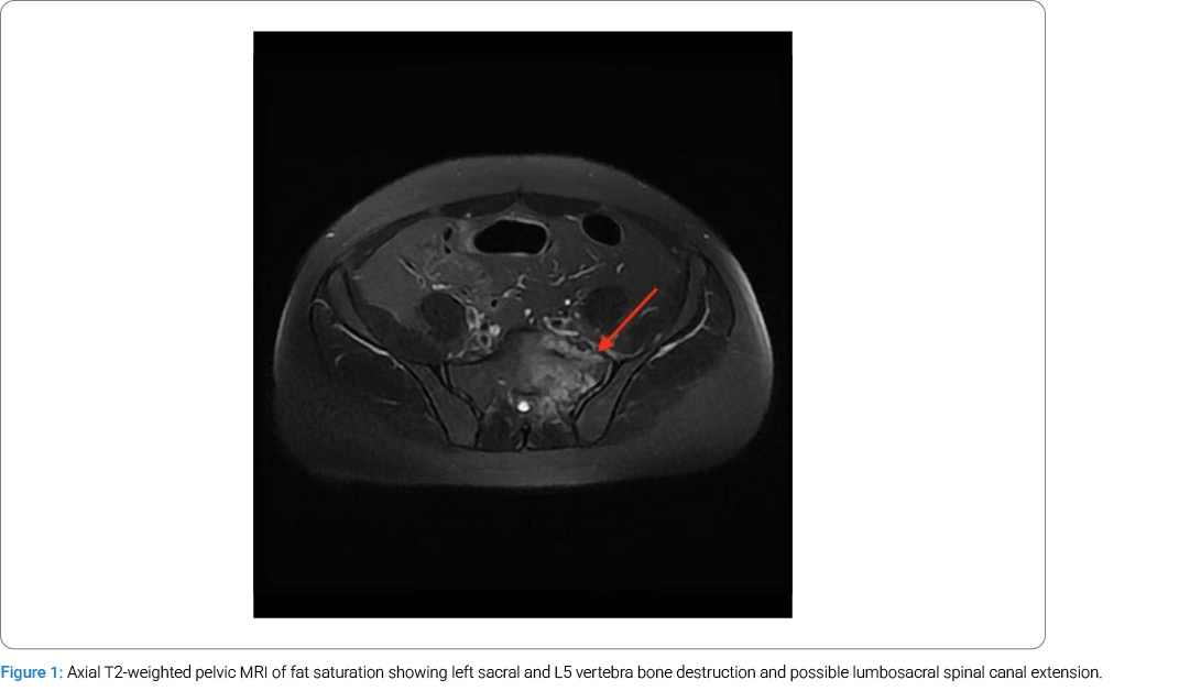
Two days later, he reported to the ED once again, with complaints of persistent headache, fatigue, myalgia, multiple episodes of vomiting, photophobia, and new-onset neck stiffness and pain radiating throughout the shoulders and spine. He was afebrile and hemodynamically stable. No focal neurologic deficits were documented, but he was noted to have weakness in bilateral upper and lower extremities. The abdomen and the midline cervical to lumbar regions were tender to palpation. Laboratory studies demonstrated leukocytosis, hyponatremia, and hypokalemia. Blood cultures were obtained. Both attempts at lumbar puncture in the ED were unsuccessful due to the patient’s pain intolerance. CXR and NC-CT of the head and cervical spine were unremarkable. Contrast-enhanced CT imaging of the abdomen and pelvis revealed abnormal osteolysis of the sacrum and left sacral ala with associated soft tissue thickening and prominent lymph nodes along the left common iliac chain. MRI of the pelvis confirmed an irregular, heterogeneous enhancing process measuring 6.3 cm, involving the lumbosacral spine (left sacrum and L5 vertebra), with associated bone destruction and possible lumbosacral spinal canal extension, as shown in (Figure 1).
At this time, the patient was admitted with suspected meningitis. Empiric intravenous ceftriaxone, vancomycin, and fluconazole were initiated. On hospital day 3, a fluoroscopy-guided lumbar puncture subsequently demonstrated cloudy Cerebrospinal Fluid (CSF) with a markedly elevated opening pressure of 62 cm H₂O, a negative meningitis panel and acid-fast bacilli smear, and no organisms on Gram stain. Cytology revealed neutrophilic predominance. Serum Coccidioides antibodies were positive with a complement fixation titer of 1:512, consistent with disseminated coccidioidomycosis.
On hospital day 6, CT-guided aspiration of the left lumbosacral lesion yielded purulent material with granulomatous inflammation and fungal elements consistent with Coccidioides, prompting initiation of intravenous amphotericin B. Repeat lumbar puncture on hospital day 8 demonstrated improvement in opening pressure (38 cm H₂O) and CSF parameters, with a coccidioidal serologic titer of 1:4.
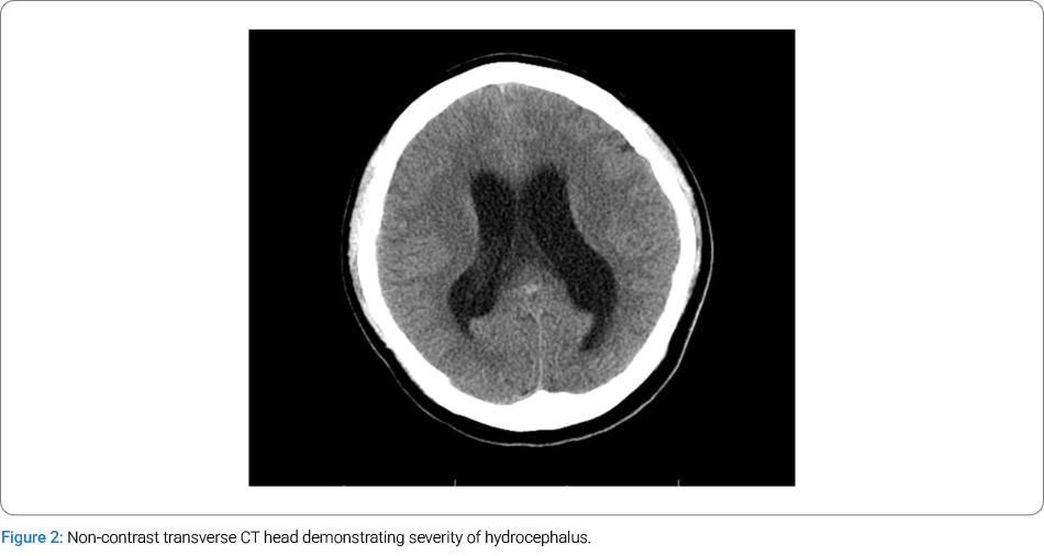
On hospital day 11, the patient developed worsening somnolence accompanied by altered mental status. NC-CT of the head demonstrated diffuse ventriculomegaly with sulcal effacement consistent with hydrocephalus (Figure 2). An External Ventricular Drain (EVD) was emergently placed with immediate drainage of CSF under elevated pressure. Pressure drainage was targeted for -5 cm H2O below External Auditory Canal (EAC) pressure. The patient was then transferred to the critical care unit where he resided for the remainder of his hospital stay. By the following day, his mental status improved and repeat NC-CT imaging showed decreased ventricular size without evidence of intracranial hemorrhage.
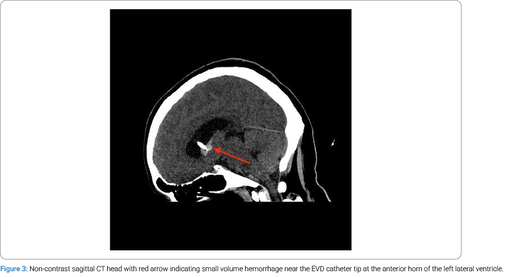
Two days after EVD placement (hospital day 13), the patient once again developed lethargy. NC-CT of the head demonstrated interval enlargement of the ventricles approximately 8 mm with a small volume intraventricular hemorrhage near the catheter tip at the anterior horn of the left lateral ventricle (Figure 3). Neurosurgery performed emergent revision of the right frontal EVD to a more lateral positioning with continued drainage goals. Initial drainage of blood-tinged CSF was noted by neurosurgery.
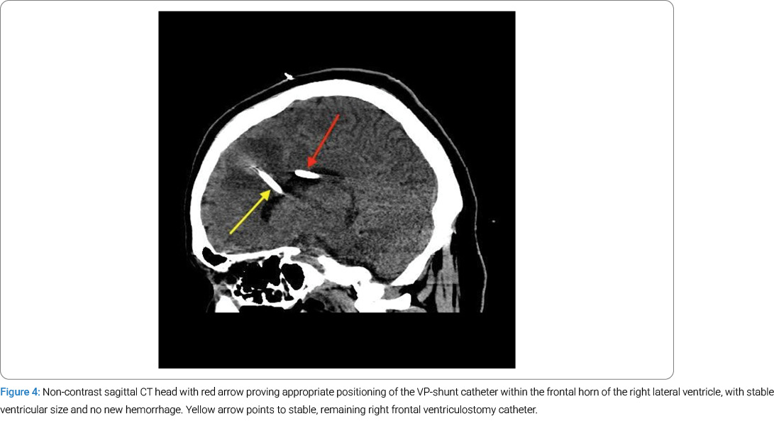
After achieving clear CSF drainage from the EVD and NC-CT on hospital day 23, demonstrating decreased pericatheter hemorrhage, a permanent Medtronic right parietal pressure-valve Ventriculoperitoneal (VP) shunt set to 0 cm H2O was placed on hospital day 24. The neurosurgical procedure was performed using a Stealth navigation Axiem system through the right parietal scalp. The distal catheter was placed intraperitoneally using laparoscopic techniques. The EVD was clamped and left in place as a precaution by neurosurgery in the event of a VP shunt malfunction. Postoperative NC-CT confirmed appropriate positioning of the shunt catheter within the frontal horn of the right lateral ventricle, with stable ventricular size and no new hemorrhage (Figure 4). For this reason, neurosurgery decided to remove the EVD on postoperative day 1 (POD-1).
On POD-5, a VP shunt tap was performed to evaluate its patency and function. Despite evidence of shunt patency on tap testing (pressure < 1 cm H2O) and maximal valve adjustment, the patient demonstrated progressive ventriculomegaly and persistent symptoms of headache and nausea. Rather than a mechanical shunt malfunction, the authors presume that the ventricular enlargement was due to inadequate CSF diversion in the setting of decreased ventricular compliance and impaired CSF absorption, necessitating greater CSF outflow than what could be achieved with the existing valve-regulated system. A shunt revision was performed to a Medtronic valveless, siphonless system incorporating a Rickham reservoir and a slit distal catheter to facilitate continuous CSF drainage. Revision was performed one week after the initial procedure, with follow-up NC-CT demonstrating decreased ventricular size and intraventricular air. Fluconazole was discontinued the following day after 30 days of therapy due to transaminase elevation three-fold above baseline. Antifungal therapy was transitioned to voriconazole reflexively with notable stabilization of transaminases following. Systemic amphotericin B was continued, with planned initiation of intrathecal amphotericin B therapy due to the severity of the disease course. It must be noted that throughout the course of the patient’s disease trajectory, the patient continued to experience headaches, nausea, and vomiting, likely attributable to amphotericin therapy and hydrocephalus in the setting of meningitis.
Over the subsequent weeks, the patient required escalating CSF diversion, ultimately necessitating bilateral frontal EVD placement, despite a previously placed VP shunt. Intraparenchymal hemorrhages, similar to prior episodes, complicated this. In the setting of clinical deterioration and ongoing complications, the patient was transferred to a higher level of care for Ommaya reservoir placement. A final NC-CT demonstrated stable positioning of ventriculostomy catheters and multicompartmental intracranial hemorrhages without new hemorrhage or progressive mass effect. He was subsequently lost to follow-up.
Discussion
This case highlights the importance of maintaining a high index of suspicion for coccidioidal meningitis in patients presenting with persistent or progressive headaches in endemic regions, even when initial imaging and laboratory findings are unrevealing, and the patient is immunocompetent. The patient’s hospital course was complicated by severe hydrocephalus requiring EVD placement, followed by VP shunt insertion with subsequent revision and additional bilateral frontal EVD placement. Before revision, the shunt was confirmed to be mechanically patent at bedside, yet progressive ventriculomegaly persisted despite the valve being set to its lowest programmable pressure of 0 cm H2O. Given that the programmable valve was already optimized to its minimum threshold, the inability to further reduce the opening pressure with standard valve mechanisms necessitated evaluating a valveless diversion system. The decision to convert to a valveless system, consisting of a Rickham reservoir and slit-end distal catheter, was driven by the need to eliminate all valve-imposed resistance to CSF drainage. This patient’s hydrocephalus evolved from an initial high-pressure state to a Low-Pressure Hydrocephalus (LPH) evidenced by neurological deterioration that occurred at intracranial pressures below the opening pressure of the shunt valve. Some diagnostic criteria for LPH include intracranial pressure ≤ 5 cm H2O and clinical improvement following subatmospheric CSF drainage [4]. Although LPH remains incompletely understood, current evidence suggests that it is multifactorial. Michael et al. describe a hysteretic pressure–volume relationship in which initial high intracranial pressures cause deformation of the brain parenchyma, and once acute pressure is relieved, ventricles fail to return to normal size even at normal or low pressures [5]. We propose that the loss of elasticity in brain parenchyma led to reduced ventricular compliance, and the granulomatous inflammation from coccidioidal meningitis resulted in fibrotic impairment of CSF absorption.
Similarly, a previous case report describes successful treatment of LPH following conversion to a valveless siphoning shunt in a patient with LPH refractory to conventional valve-based systems [5]. These complications underscore the substantial morbidity associated with central nervous system dissemination of Coccidioides. In addition, patients diagnosed with coccidioidal meningitis often require lifelong antifungal therapy and close long-term follow-up due to the high risk of relapse.
Coccidioidomycosis remains diagnostically challenging, with an enhanced surveillance study of 186 patients demonstrating a median interval of 38 days from initial healthcare presentation to confirmed diagnosis [6].
Prior studies highlight the complexity of managing coccidioidal meningitis. In a 10-year retrospective cohort of 133 patients, 60% required VP shunt placement for intracranial hypertension, and nearly half of those required revision due to shunt failure, underscoring the high rate of complications associated with CSF diversion. Additionally, the same study reported rates of antifungal nonadherence at 43% and mortality in 23% due to complications at an average of 22 months following diagnosis, further emphasizing the challenges in achieving sustained disease control [7].
Similarly, a retrospective analysis of 44 patients with coccidioidomycosis-associated hydrocephalus demonstrated a predominance in male patients of African or Asian descent and identified mechanical dysfunction as the leading cause of shunt failure, accounting for 81% of revisions. Notably, patients underwent an average of 2.5 shunt revisions over the course of their disease, underscoring the difficulty in achieving durable CSF diversion in this population [8]. Our patient, who is a male of Asian descent, reflects this reported demographic pattern and experienced a similarly complex neurosurgical course.
CNS dissemination of coccidioidomycosis is an uncommon but severe manifestation, occurring more frequently in immunocompromised individuals; however, cases in immunocompetent hosts are well documented. A systematic review of adult cases published between 2012 and 2022 identified 14 individual cases of coccidioidal meningitis, most commonly affecting male, HIV-negative patients residing in endemic regions [9]. Our patient, an immunocompetent Asian male, reflects this demographic pattern, showing the potential for aggressive CNS involvement despite preserved immune function.
Skeletal coccidioidomycosis arises from hematogenous dissemination and primarily involves the axial skeleton, leading to progressive bone destruction. Clinical manifestations are variable and include osteomyelitis, osteolysis, and discitis, with symptoms such as back or neck pain, height loss, lower extremity weakness, and constitutional symptoms. Pulmonary findings are often absent. There is also a risk of hematogenous spread of vertebral involvement to the skull, affecting the pachymeninges and subarachnoid space [10]. This case illustrates how disseminated coccidioidomycosis can present insidiously in an immunocompetent host, with vertebral involvement progressing to fulminant CNS disease.
Conclusion
Coccidioidomycosis is most often seen as a respiratory illness that may propagate into systemic illness. Because of this generality, physicians often rely on abnormal pulmonary findings to gauge investigation into Coccidioides spp. infection without discerning other bodily areas of origin. We suspect that the patient’s lumbosacral osteolysis was the source of his disseminated coccidioidomycosis given the clinical picture, imaging results, laboratory findings, and cytologic/pathologic findings. This unique case sheds light on the complexities and dangers of Coccidioides spp. in infecting even the most immunocompetent individuals.
Human Ethics
Consent was obtained or waived by all participants in this study.
Conflict of Interest
The authors declare no potential conflicts of interest with respect to the research, authorship, and/or publication of this article. Informed consent was obtained for this publication.
Cite this article
Aghakhani N, Tailor C, Bajwa MR. Disseminated Coccidioidal Meningitis in a Previously Healthy Adult Complicated by Hydrocephalus Requiring Ventriculoperitoneal Shunt Placement: A Case Report. Clin Case Rep J. 2026;7(5):1–5.
Keywords
Disseminated coccidioides, meningitis, coccidioides meningitis, Valley fever meningitis, coccidioidomycosis
Copyright
© 2026 Nina Aghakhani. This is an open access article distributed under the terms of the Creative Commons Attribution 4.0 International License (CC BY-4.0).